
Make it
Yours.
Yours.
Edit, remix and personalize with your own text
Or start from these designs
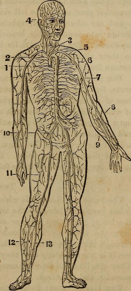
Identifier: anatomyphysiolog00jord (find matches)Title: "Anatomy, physiology and laws of health;"Year: 1885 (1880s)Authors: Jordan, Johnson H. (from old catalog)Subjects: Medicine, Popular Anatomy PhysiologyPublisher: Chicago, W. H. Moore & co.Contributing Library: The Library of CongressDigitizing Sponsor: The Library of CongressView Book Page: Book ViewerAbout This Book: Catalog EntryView All Images: All Images From BookClick here to view book online to see this illustration in context in a browseable online version of this book.Text Appearing Before Image:ng it freely. The arteries are enveloped in sheaths of a loose cellular texture(the same which envelop the muscles), which separate them fromthe adjacent parts, and also enclose the veins and nerves which generally accompany them. All the larger arteries are deeply seated, by which arrangementthey are protected from injury by accidents, while the veins, whichdo not involve so serious consequences in case of wounds, are gene-rally placed near the surface of the body—often immediately underthe skin, as on the back of the hand, and upon the wrist. Fig. 6. Fig. 6.—The Arterial System :— 1. Commencement of the aorta, where it leaves the heart. 2. Arch of the aorta. 3. Carotid artery— (one on each side). 4. Temporal artery. 5. Subclavian artery. 6. Axillary, artery. 7. Brachial artery. 8. Eadial artery. 9. Ulnar artery. 10. Iliac artery. 11. Femoral artery. 12. Tibial artery. 13. Peroneal artery. H^g^All of these arteries are in pairs; thatis, one on each side, or in each extremity.Text Appearing After Image:The Arterial System.The Aorta, which conveys the pure blood to all parts of the body,proceeds from the left ventricle of the heart, rises toward the left 28 ANATOMY, PHYSIOLOGY-AND clavicle or collar bone, and turns in the form of an arch toward theback and left side, and passes down behind the heart, through thediaphragm, along the spine, sending off numberless branches—whichalso divide and subdivide, like the branches of a tree—to all theinternal organs and parts of the body, and finally, in the lower partof the abdominal cavity, it bifurcates—that is, divides into two mainbranches, one passing down each leg, constantly sending off branches,till the whole terminate in what are called capillaries—small blood-vessels too delicate to be seen distinctly without the aid of a micro-scope, and which will be described presently. From the top of the arch of the aorta three main branches go off.The first, or the one on the right, soon divides, a branch going to theright arm—the righNote About ImagesPlease note that these images are extracted from scanned page images that may have been digitally enhanced for readability - coloration and appearance of these illustrations may not perfectly resemble the original work.
Original public domain image from Wikimedia Commons
Public DomainFree CC0 image for Personal and Business use
MonthlyYearlySave 50%
Get Premium
Professional design resources and creative tools
from
from
$
6.50
/mo
$78 billed yearly
- Unlimited downloads
- Ad-free experience
- Unlock millions of creative assets and our entire Creative Studio of editable templates, mockups and design tools