https://creativecommons.org/publicdomain/zero/1.0/https://www.rawpixel.com/image/8718004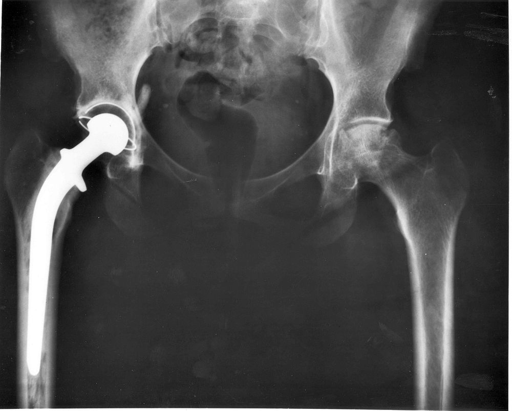
An A-P X-ray of a pelvis showing a total hip joint replacement. The right hip joint (on the left in the photograph) has been replaced. A metal prostheses is cemented in the top of the right femur and the head of the femur has been replaced by the rounded head of the prosthesis. A white plastic cup is cemented into the acetabulum to complete the two surfaces of the artificial "ball and socket" joint.Although not the case here, hip prostheses can also be made of a ceramic material which rarely wears out during the patient's lifetime. During the operation, a bonding cement is used to fix the metal prostheses into the shaft of the femur and the plastic cup to the acetabulum (socket in the hip bone). One of the leading reasons for hip replacement is osteoarthritis of the hip joint in which virtually all of the cartilage around the top of the femur bone deteriorates due to wear over time, leaving a grinding bone-on-bone situation with the bone surfaces becoming roughened leading to pain and stiffness. Narrowing of joint space (the space between the acetabulum and the head of the femur) is also a feature of osteoarthritis. There may be other changes which are not entirely clear on this A-P X-ray, which probably should be reported together with lateral X-rays of the hips, or with modern computerised imaging techniques.Keywords: total hip replacement, prosthesis, osteoarthritis, X-ray.
Original public domain image from Wikimedia Commons
Public DomainFree CC0 image for Personal and Business use