https://creativecommons.org/publicdomain/zero/1.0/https://www.rawpixel.com/image/9974046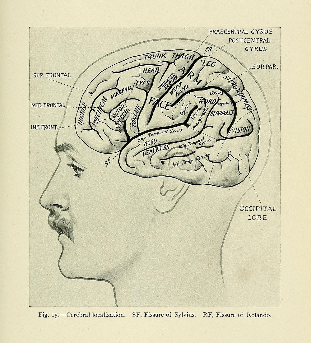
Identifier: textbookofclinic00eise (find matches)Title: A text-book of clinical anatomy : for students and practitionersYear: 1907 (1900s)Authors: Eisendrath, Daniel N. (Daniel Nathan), b. 1867Subjects: Human anatomy AnatomyPublisher: Philadelphia London : W.B. Saunders CompanyContributing Library: Francis A. Countway Library of MedicineDigitizing Sponsor: Open Knowledge Commons and Harvard Medical SchoolView Book Page: Book ViewerAbout This Book: Catalog EntryView All Images: All Images From BookClick here to view book online to see this illustration in context in a browseable online version of this book.Text Appearing Before Image:tionsof brain, and of middle meningeal artery to skull. M.A., Anterior or ascending branch ofthe middle meningeal artery. M.P., Posterior or horizontal branch of the middle menin-geal artery. These two join so that the main trunk lies about opposite the middle of thezygoma. F.R., Fissure of Rolando, lying between 4, the ascending frontal, and 5, theascending parietal convolution. F.S., Fissure of Sylvius, that is, the horizontal limb. 1,2, and 3, First, second, and third frontal convolution. 6, Parietal lobe. 7, Occipital lobe.8, 9, and 10, First, second, and third temporal convolution. L.S., Lateral sinus. Thedotted line shows that portion of the lateral sinus which descends on the inner side of themastoid process (sigmoid sinus). The remainder of the lateral sinus is shown passingsomewhat horizontally backward beneath the occipital lobe (7). A.M., Projection ofmastoid antrum, on surface. 5i ,PRAECENTRAL GYRUS.POSTCENTRALFR y GYRUS WJ5UEPAR,; SUP. FRONTAL i MID.FRONTAL. INF. FRONT. -Text Appearing After Image:OCCIPITALLOBE Fig. 15.--Cerebral localization. SF, Fissure of Sylvius. RF, Fissure of Rolando. 53Note About ImagesPlease note that these images are extracted from scanned page images that may have been digitally enhanced for readability - coloration and appearance of these illustrations may not perfectly resemble the original work.
Original public domain image from Wikimedia Commons
Public DomainFree CC0 image for Personal and Business use