Or start from these designs
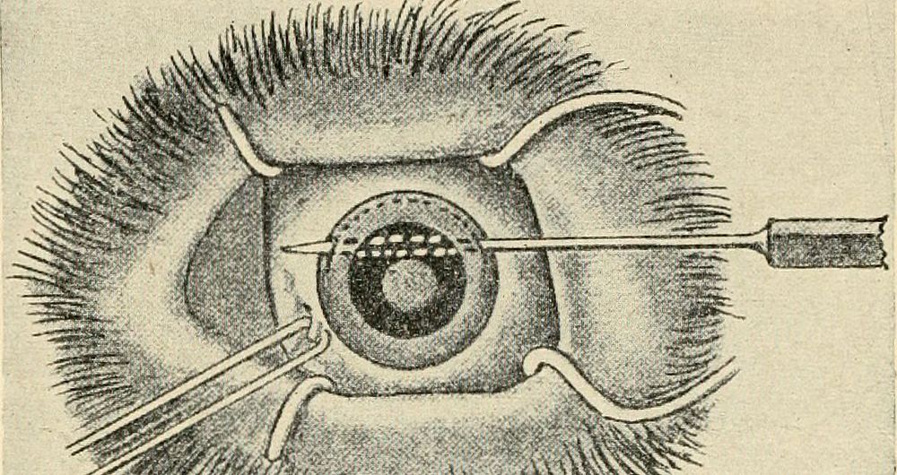
Identifier: diseasesofdogthe00ml (find matches)Title: Diseases of the dog and their treatmentYear: 1911 (1910s)Authors: Müller, Georg Alfred, 1851-1923 Glass, AlexanderSubjects: Horses Dogs -- DiseasesPublisher: Chicago, Ill. : Alexander EgerContributing Library: Webster Family Library of Veterinary MedicineDigitizing Sponsor: Tufts UniversityView Book Page: Book ViewerAbout This Book: Catalog EntryView All Images: All Images From BookClick here to view book online to see this illustration in context in a browseable online version of this book.Text Appearing Before Image:sion. Linear Extraction.—This operation is used where there is completeopacity of the lens and enlargement of the lens and contraction of the 420 DISEASES OF THE EYES anterior chamber. After liaving prepared the dog as in the operationfor discission, an assistant holding the eyelids apart, with anotherforceps seize the conjunctiva of the eyeball in the neighborhoodof the median line of the eyeball, at the same time everting the uppereyelid. We then make an incision by means of Graefes cataract knife(Fig. 143, b), about 5 mm. broad, through the cornea, about 2 or 3 mm.from the border of the sclerotic membrane. We then pass a discissionneedle through the wound, split open the lenticular capsule, as in discis-sion, and empty the soft parts of the cataract by means of Davielsspoon (Fig. 143, c). Any remnants of the cut capsule which may notbe removed at the time are left to be reabsorbed. If during the operationAve observe prolapsus of the iris, we must try to restore it to its positionText Appearing After Image:■%//#fMii;m# Fig. 145.—Lobular extraction of leii.s. (Cadiot and Breton.) by means of Daviels spoon (Fig. 143, c). If this is not possible, wemay cut it off close to the wound of the cornea. It is very evident that linear extraction is only to be performed incases of complete softening of the lens. This may be recognized liytotal opacity of the lens and alteration of the iris, and also when theanterior ca))sule is pushed toward the cornea. Lobular Extraction.—Lobular extraction is indicated in hard cata-ract, which is generally senile, where the lens may be prolapsed into theanterior chamber and where discission will only produce an imperfectresult—that is to say, where reabsorption of the lens does not progressproperly. It is performed in the following manner: Make an incision into the cornea exactly as in linear extraction, bymeans of Graefes cataract knife, but it must be enlarged to 8 or 10 mm,(Fig. 145). After that the capsule of the lens is split by the discissionneedle,Note About ImagesPlease note that these images are extracted from scanned page images that may have been digitally enhanced for readability - coloration and appearance of these illustrations may not perfectly resemble the original work.
Original public domain image from Wikimedia Commons
Public DomainFree CC0 image for Personal and Business use
MonthlyYearlySave 50%
Get Premium
Professional design resources and creative tools
from
from
$
6.50
/mo
$78 billed yearly
- Unlimited downloads
- Ad-free experience
- Unlock millions of creative assets and our entire Creative Studio of editable templates, mockups and design tools