Make it
Yours.
Yours.
Edit, remix and personalize with your own text
Or start from these designs
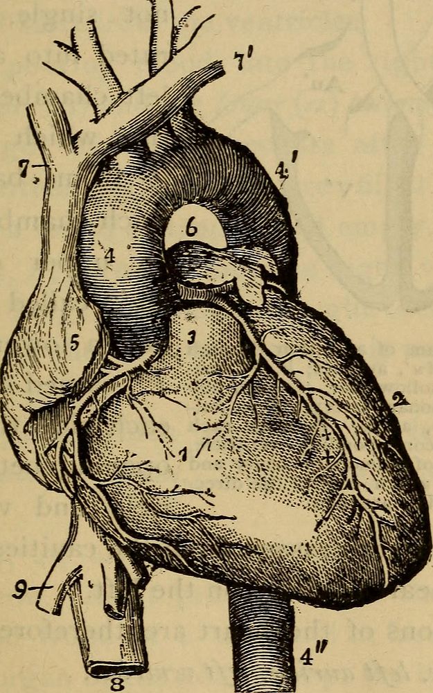
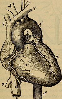
Identifier: humanbodybeginne00mart (find matches)Title: The human body. A beginner's text-book of anatomy, physiology and hygiene ..Year: 1884 (1880s)Authors: Martin, H. Newell (Henry Newell), 1848-1896 Martin, Hetty Cary, (from old catalog) joint authorSubjects: PhysiologyPublisher: New York, H. Holt and companyContributing Library: The Library of CongressDigitizing Sponsor: The Library of CongressView Book Page: Book ViewerAbout This Book: Catalog EntryView All Images: All Images From BookClick here to view book online to see this illustration in context in a browseable online version of this book.Text Appearing Before Image:healthy living blood-vessels, no fibrin forms init, and it does not clot. But as soon as blood gets out-side of the vessels, or whenever their lining is injured,clotting takes place. In this way, the ends of thesmall blood-vessels in a cut finger are soon clogged up,if we can only stop the flow for a little and give timefor a clot to form in them. 11. What is the consistency of fresh blood? What changes occurin it during the first five or six minutes after it is drawn ? What isthe solidifying of the blood called ? To what is it due ? What isserum ? What is the clot ? 12. Use of coagulation ? When does it not occur ? When does ittake place? Why does a cut finger stop bleeding after a short time? THE HEART. 139 13. The Heart (Fig. 37) resembles a pear in form, andis placed in a slanting position inside the chest, with itssmaller end downwards. It lies just above the diaphragm(Fig. 2), and behind the lower two-thirds of the breast-bone. Its upper end, or base (so called because it is theText Appearing After Image:Fig. 37.—The heart and the arteries and veins opening into it, seen from thefront. The pulmonary artery has been cut short close to its beginning, i, rightventricle; 2. left ventricle; 3, root of the pulmonary artery; 4, 4, ^\ the aorta; 5,part of the right auricle; 6, part of the left auricle; 7, 7, innominate veins joiningthe upper vena cava; 8, inferior vena cava; 9, one of the veins from the liver, join-ing the inferior vena cava. larger end, although the upper), projects a little to theright of that bone, and its lower end, or apex, a littleto the left, where it may easily be felt beating by pressingwith the finger between the cartilages (p. i8) of the 13. Shape and position of the heart ? Where does its base project ?Where may it§ apex be felt beating? Its size ? I40 THE PERICARDIUM,Note About ImagesPlease note that these images are extracted from scanned page images that may have been digitally enhanced for readability - coloration and appearance of these illustrations may not perfectly resemble the original work.
Original public domain image from Wikimedia Commons
Public DomainFree CC0 image for Personal and Business use
MonthlyYearlySave 50%
Get Premium
Professional design resources and creative tools
from
from
$
6.50
/mo
$78 billed yearly
- Unlimited downloads
- Ad-free experience
- Unlock millions of creative assets and our entire Creative Studio of editable templates, mockups and design tools